Pattern definitions
Reference definitions for every annotation pattern, grouped by category and ordered as on the analysis page. ← Back to instructions
| Pattern | Definition |
|---|---|
| TOCO |
No definition yet.
|
| Uterine contraction |
Uterine contraction — a transient increase in uterine tone recorded on the tocograph (TOCO), rising from the baseline uterine tone and returning to it.
For annotation, only the start (the trace leaving the baseline TOCO) and the end (its return to the baseline TOCO tone) are marked — the peak is not annotated. [1] |
| Baseline TOCO |
Baseline TOCO — the stretches of the toco trace between contractions, representing the resting uterine tone. With external tocodynamometry only a relative value is obtained (it drifts with sensor and patient movement), so the absolute basal tone cannot be read reliably — internal intra-uterine pressure monitoring is required for that.
[1] |
| Artifact |
Artifact (TOCO) — something appears on the toco channel that is not a uterine contraction (e.g. maternal movement, repositioning of the sensor, coughing or pushing on the transducer).
|
| Poorly captured |
The TOCO tracing is too poor to locate the uterine contractions: it is impossible to identify individual contractions over this period (weak signal, displaced probe, excessive noise...). Different from an artifact, which is something appearing on the TOCO channel that is not a contraction.
|
| Not analysed/Unsure |
Not analysed / Unsure (TOCO) — a segment of the toco channel left unannotated, or where the annotator is unsure whether it is a contraction, baseline tone or artefact. Such periods are excluded from the ground truth for this channel (they do not count in training/evaluating the IA)
|
| MHR |
MHR (maternal heart rate) — the channel of the mother's own heart rate, acquired by a dedicated sensor (maternal pulse oximeter or ECG with the TOCO Belt). Marking it as true or false signal helps recognise when a Doppler FHR sensor has locked onto the maternal pulse instead of the fetal one.
An important dataset has already been labeled for this category. This is not necessary to annotate this part unless the recording is interesting. |
| True signal |
True signal (MHR) — a portion of the maternal-rate channel that faithfully reflects approximately the true maternal heart rate. During labour it usually lies below the FHR, rises with contractions and maternal pushing, and may show accelerations coinciding with expulsive efforts.
|
| False signal |
False signal (MHR channel) — a portion of the trace annotated on the maternal-rate channel that does not reflect the true maternal heart rate. It arises from the same causes as FHR false signals: the sensor may report a harmonic (double, half, triple) of the MHR or may lose/ambiguously capture the signal.
A false continuous line interpolating true and false signals over a period of 15s is technically possible. 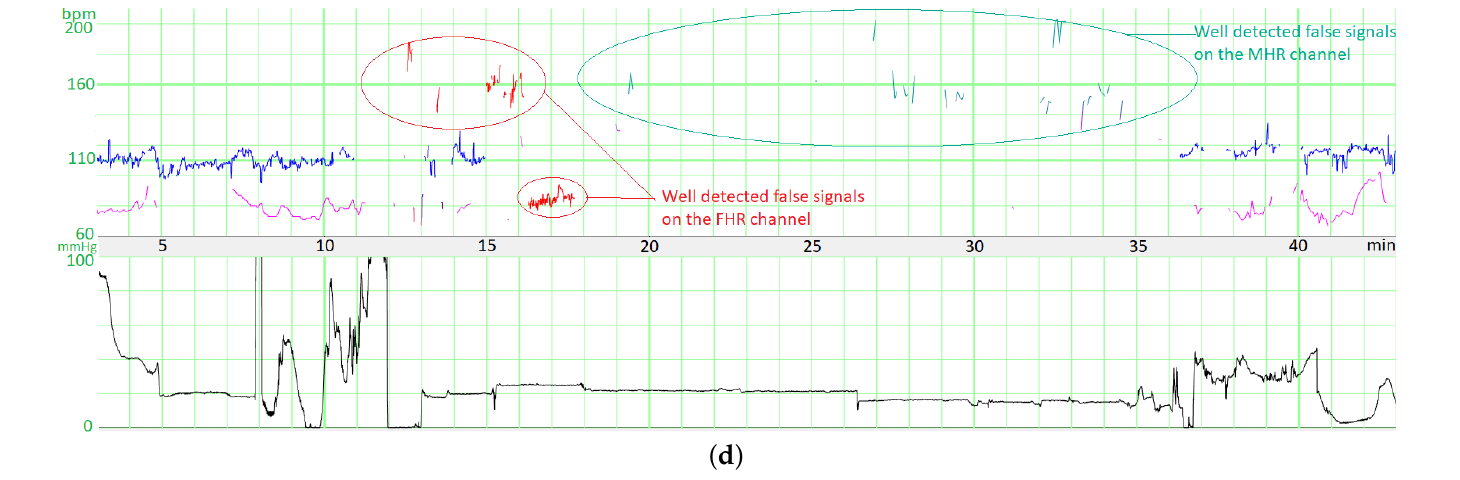 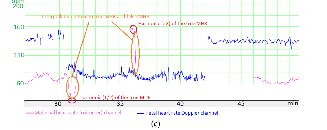 [1] |
| Not analysed/Unsure |
Not analysed / Unsure (MHR) — a segment of the maternal-rate channel left unannotated, or where it is uncertain whether the trace is a true or a false maternal signal. Excluded from the ground truth for this channel.
|
| FHR |
FHR (fetal heart rate) channel — a template applied to each available fetal-heart-rate source: FHR1 (external Doppler / ultrasound) and FHR2 (internal scalp ECG). Every FHR annotation (true/false signal, then the morphological and variability analysis) is made independently on each channel.
An important dataset has already been labeled for this category. This is not necessary to annotate this part unless the recording is interesting. |
| FHR - False/True |
FHR — False vs True signal — the first decision on each FHR channel: does the trace reflect the true fetal heart rate (true signal) or not (false signal)? Morphological and variability analysis is performed only on segments annotated as true signal. The external Doppler channel is markedly more prone to false signal than the internal scalp-ECG channel.
[1] |
| True signal |
True signal (FHR) — a portion of the FHR trace that faithfully reflects the true fetal heart rate. Only these segments undergo morphological analysis (baseline, accelerations, decelerations) and variability assessment. On the Doppler channel a true signal must be distinguished from maternal-rate pick-up and from harmonics (double or half the true rate).
[1] |
| False Signal |
False signal (FHR) — a portion of the FHR trace that does not reflect the true fetal heart rate. On a Doppler (ultrasound) sensor this is frequent, especially during the second stage of labour, and can be dangerous when it masks fetal distress.
Main mechanisms (Boudet et al. 2022): Maternal heart rate (MHR) recorded instead of the fetal rate — the sensor locks onto the maternal pulse. During the second stage of labor, one should be very cautious of the “double mountain peak” sign when large amplitude accelerations coincide with ongoing uterine contractions (Figure below) because of erroneous monitoring of the maternal heart rate as the FHR. 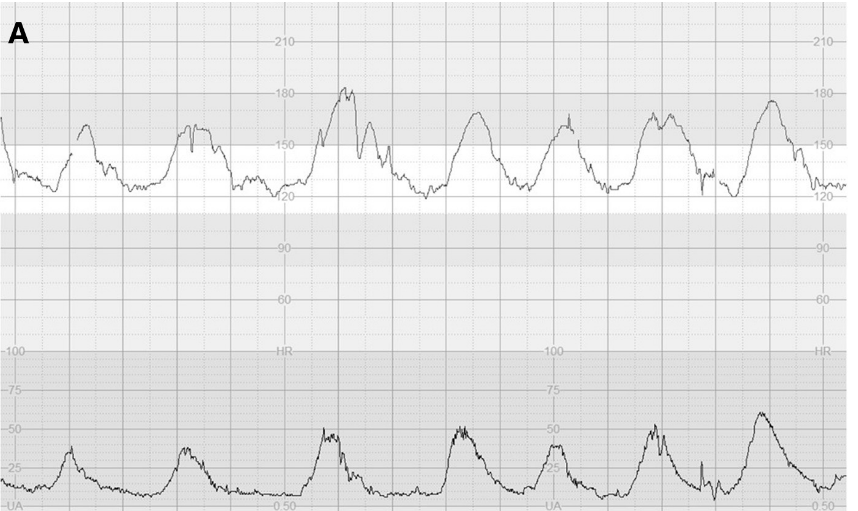 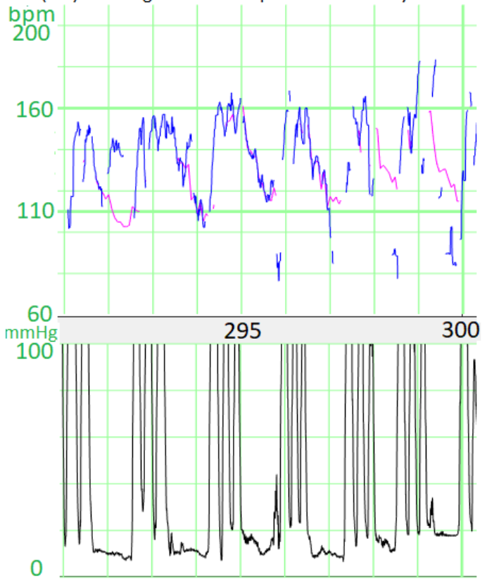 Harmonics of the true rate — the sensor reports double, half or (more rarely) triple the true FHR (or a harmonic of the MHR). Missing / ambiguous signal — dropout or an unstable, uninterpretable trace. Generally, there are missing signal periods between false signals period and true signal periods, but sometimes it can be interpolated over a few seconds. 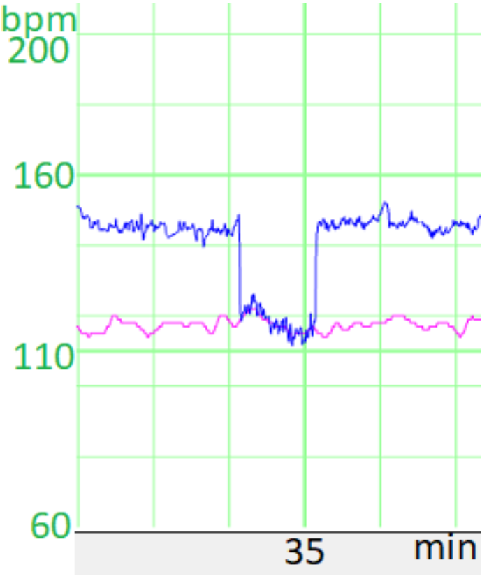 The internal (scalp-ECG) channel is far less prone to false signals, but it can still have aberrant signals. [1] [2] |
| Not analysed/Unsure |
Not analysed / Unsure (FHR true/false) — a segment of an FHR channel left unannotated, or where it is uncertain whether the trace is a true or a false signal. Excluded from the ground truth for this channel.
|
| Morphological Analysis |
Morphological analysis — the description of the shape of the FHR over time on a true-signal segment: the baseline level and the periodic / episodic events superimposed on it (accelerations and the different types of deceleration). It is performed only where the FHR is a true signal.
|
| Baseline period |
Baseline (FHR) — the mean level of the FHR when it is stable, read over segments of at least 2 minutes (excluding accelerations, decelerations and marked-variability periods).
The Baseline period annotation marks the stretches of trace that represent this stable baseline (i.e. neither acceleration nor deceleration). |
| Neither Baseline nor A/D |
Neither baseline nor A/D — a fluctuation that is neither a stable baseline nor a true acceleration/deceleration, typically a small acceleration or deceleration that does not reach the 15 bpm / 15 s criteria.
|
| Deceleration |
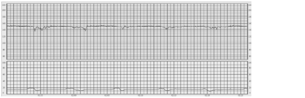
FIGO Definition: Decreases in the FHR below the baseline, of more than 15 bpm in amplitude, and lasting more than 15 seconds.
|
| Not analysed/Unsure type |
Deceleration — unsure / not analysed type — a period recognised as a deceleration but whose sub-type (early, variable, late, prolonged) has not yet been determined or is uncertain.
|
| Early |
FIGO Definition: decelerations that are shallow, short-lasting, with normal variability within the deceleration and are coincident with contractions. They are believed to be caused by fetal head compression [25] and do not indicate fetal hypoxia/acidosis
|
| Variable |
FIGO Definition (2015): V-shaped deceleration with a rapid drop (onset to nadir in < 30 s), good variability within the deceleration, rapid recovery to the baseline, and varying size, shape and relationship to uterine contractions. Variable decelerations constitute the majority of decelerations during labour and reflect a response to increased arterial pressure, as occurs with umbilical cord compression and resultant transient hypoxaemia - not hypoxia/acidosis. The intervening baseline and variability should be assessed to judge central-organ oxygenation.
[1] |
| Late with normal variability |
FIGO Definition: (U-shaped and/or with reduced variability): decelerations with a gradual onset and/or a gradual return to the baseline. Gradual onset and return occurs when more than 30 seconds elapses between the beginning/end of a deceleration and its nadir. When contractions are adequately monitored, late decelerations start more than 20 seconds after the onset of a contraction, have a nadir after the acme, and a return to the baseline after the end of the contraction.
These decelerations are indicative of a chemoreceptor-mediated response to fetal hypoxemia. In the presence of a tracing with no accelerations and reduced variability, the definition of late decelerations also includes those with an amplitude of 10−15 bpm. 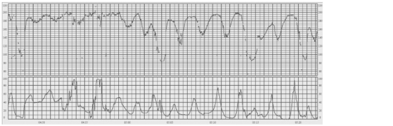 [1] |
| Late with reduced variability |
FIGO Definition: (U-shaped and/or with reduced variability): decelerations with a gradual onset and/or a gradual return to the baseline and/or reduced variability within the deceleration. Gradual onset and return occurs when more than 30 seconds elapses between the beginning/end of a deceleration and its nadir. When contractions are adequately monitored, late decelerations start more than 20 seconds after the onset of a contraction, have a nadir after the acme, and a return to the baseline after the end of the contraction.
These decelerations are indicative of a chemoreceptor-mediated response to fetal hypoxemia. In the presence of a tracing with no accelerations and reduced variability, the definition of late decelerations also includes those with an amplitude of 10−15 bpm. |
| Prolonged with normal variability |
FIGO Definition: lasting more than 3 minutes. These are likely to include a chemoreceptor-mediated component and thus to indicate hypoxemia. Decelerations exceeding 5 minutes, with FHR maintained at less than 80 bpm and reduced variability within the deceleration, are frequently associated with acute fetal hypoxia/acidosis and require emergent intervention.
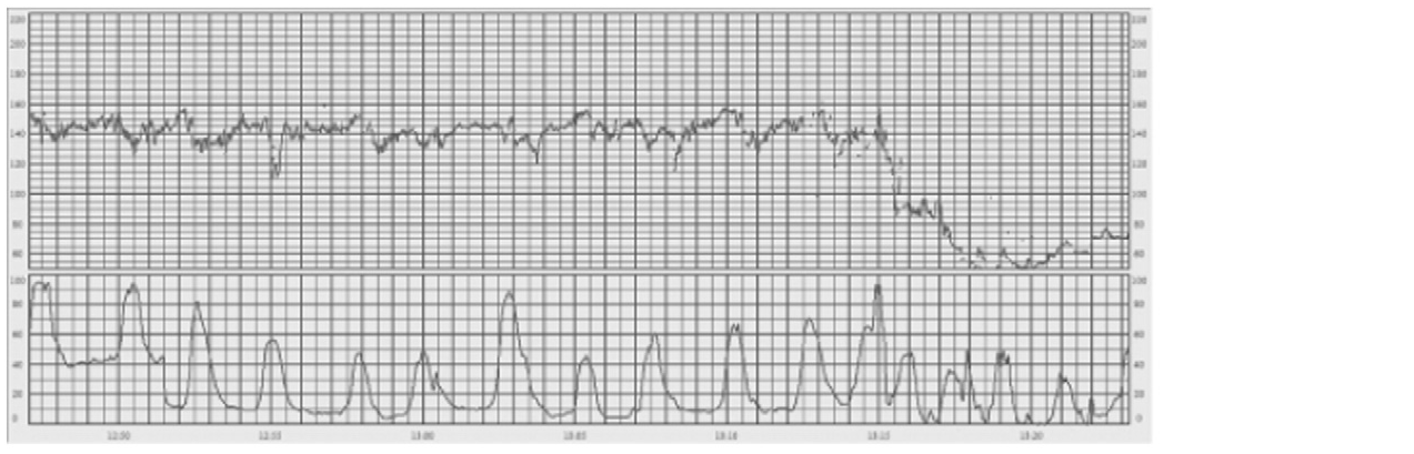 [1] |
| Prolonged with reduced variability |
FIGO Definition: lasting more than 3 minutes. These are likely to include a chemoreceptor-mediated component and thus to indicate hypoxemia. Decelerations exceeding 5 minutes, with FHR maintained at less than 80 bpm and reduced variability within the deceleration, are frequently associated with acute fetal hypoxia/acidosis and require emergent intervention.
|
| Acceleration |
FIGO Definition: Abrupt (onset to peak in less than 30 seconds) increases in FHR above the baseline, of more than 15 bpm in amplitude, and lasting more than 15 seconds but less than 10 minutes.
|
| Not analysed/Unsure type |
Acceleration for which it is unsure whether it is spurious or a true acceleration, or this detail is not analysed.
|
| True acceleration |
True acceleration. Abrupt, transient increase of the FHR >=15 bpm lasting >=15 s (but <10 min), arising from a stable, normal baseline and returning to the same baseline, with normal baseline variability. It must not be part of a deceleration. Before 32 weeks, >=10 bpm / >=10 s may suffice.
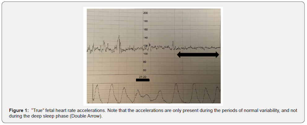 [1] |
| Shoulders (overshoot / sloping cliff) |
Shoulders (grouping overshoot and sloping cliff) - spurious accelerations that are an integral part of a deceleration, not stand-alone accelerations from a stable baseline.
How to identify: Shoulder of a variable deceleration: a rise immediately before and/or after a variable deceleration, of roughly equal amplitude on both sides. 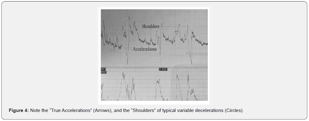 Overshoot: an exaggerated, wider, abrupt rise on the recovery (ascending) limb of a variable deceleration, larger than a true acceleration. 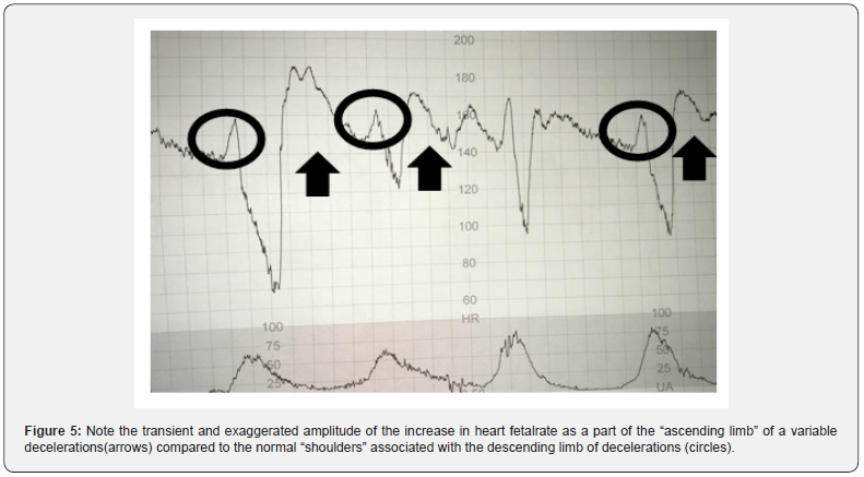 Sloping cliff sign: a shallow, gradual rise immediately preceding a shallow deceleration, with reduced baseline variability (it masks the underlying shallow deceleration). [1] |
| Not analysed/Unsure |
Not analysed / Unsure (morphology) — a true-signal FHR segment whose morphology (baseline vs acceleration vs which deceleration) has not been annotated or is uncertain. Excluded from the morphological ground truth.
|
| Variability |
FIGO Definition: This refers to the oscillations in the FHR signal, evaluated as the average bandwidth amplitude of the signal in 1-minute segments
Precision: Average over minutes not average within the minute. Annoter les périodes d'au moins 5-10 minutes de variabilité reduced, ne pas couper la période à chaque acceleration/ralentissement. Si le manque de variabilié est uniquement pendant les ralentissements, il faut les coder dans late deceleration et non ici. |
| Normal |
Normal variability — a bandwidth amplitude of 5–25 bpm (FIGO 2015).
[1] |
| Not pseudo sinusoidal pattern |
Normal variability — not pseudo-sinusoidal — ordinary normal baseline variability (bandwidth 5–25 bpm) that does not adopt a pseudo-sinusoidal shape. This is the default sub-type of normal variability, separated from the pseudo-sinusoidal pattern, which transiently mimics a sinusoidal wave in a benign context.
|
| Pseudo sinusoidal pattern |
Pseudo-sinusoidal pattern — a pattern resembling the sinusoidal pattern but with a more jagged, “saw-tooth” appearance rather than a smooth sine wave. Its duration seldom exceeds 30 minutes and it is characterised by normal patterns before and after. It has been described after maternal analgesic administration and during fetal sucking / mouth movements. It can be difficult to distinguish from a true sinusoidal pattern, the short duration being the most useful discriminator.
The typical / non-pseudo distinction rests largely on the clinical association; pseudo-sinusoidal is not the same as a saltatory pattern (cf. issue #11). 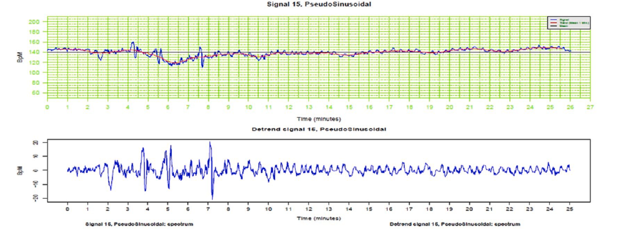 [1] |
| Reduced | |
| Unsure type |
Reduced variability — unsure type — a period of reduced baseline variability (bandwidth < 5 bpm) whose cause has not been settled: physiological deep sleep, pathological (hypoxic) reduction, or absent variability.
|
| Deep sleep/physiological |
FIGO Definition: During deep sleep, variability is usually in the lower range of normality, but the bandwidth amplitude is seldom under 5 bpm. Deep sleep can last up to 50 minutes and is associated with a stable baseline, very rare accelerations, and borderline variability.
Note: This is also called Pattern A in the literature In this category, we also put physiological reduced variability, for example for patients of sub-Saharan origin. If you are not sure just put "unsure type" instead |
| Pathologic reduced variability |
FIGO Definition: more than 50 minutes in baseline segments. Reduced variability can occur due to central nervous system hypoxia/acidosis and resulting decreased sympathetic and parasympathetic activity, but it can also be due to previous cerebral injury, infection, administration of central nervous system depressants or parasympathetic blockers. [...] Following an initially normal CTG, reduced variability due to hypoxia is very unlikely to occur during labor without preceding or concomitant decelerations and a rise in the baseline.
|
| Absent variability |
(silent pattern) Totally absent variability and reactivity, with or without decelerations and bradycardia. Absent variability and pre-terminal pattern not classified by FIGO, but constitute a fourth CTG class (pre-terminal CTG) in the STAN CTG classification system
|
| Zigzag |
Zigzag pattern.
An abrupt and erratic up-and-down fluctuation of the baseline FHR variability exceeding 25 bpm, irregular in both frequency and morphology, lasting at least 1 minute. Unlike the saltatory pattern it is transient and not uniform. 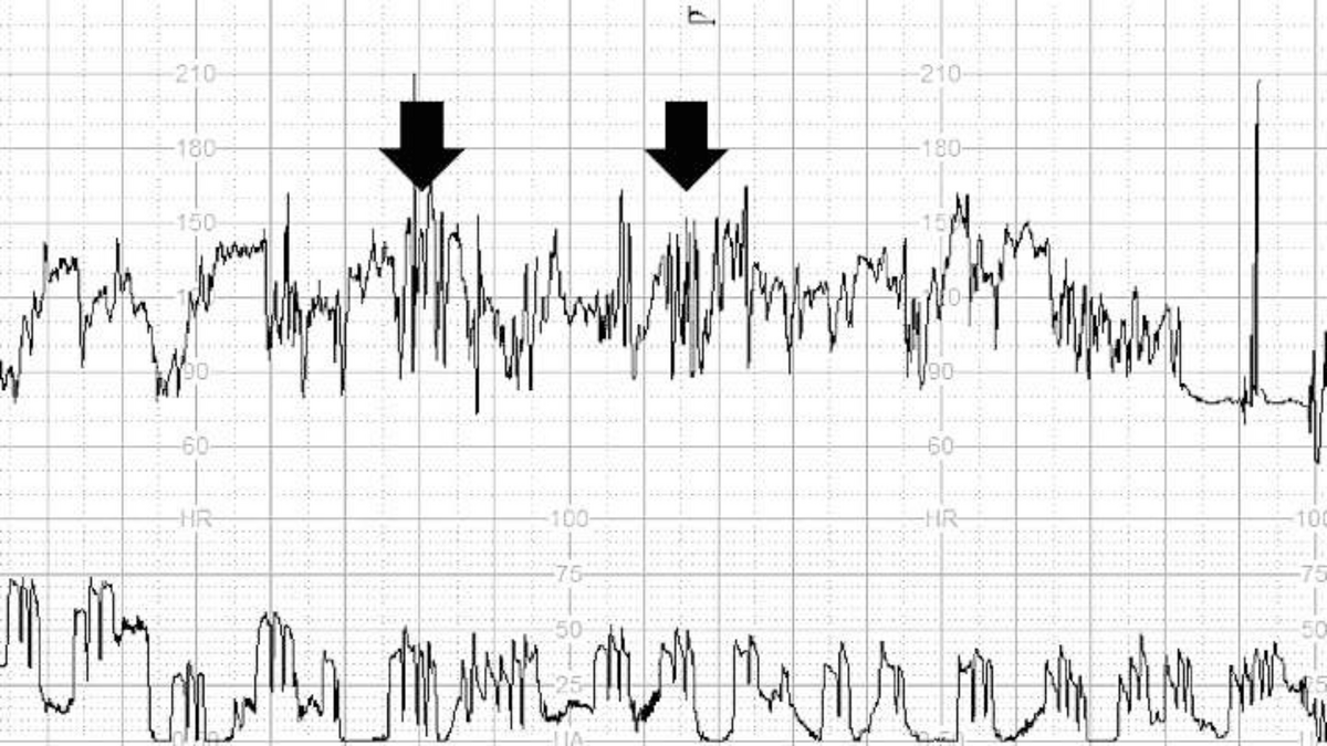 It occurs during labour when the intensity of the hypoxic stress increases with insufficient time at the baseline to ensure adequate gas exchange (rapidly evolving hypoxia). The mechanism is an autonomic instability, predominantly mediated by the parasympathetic nervous system. 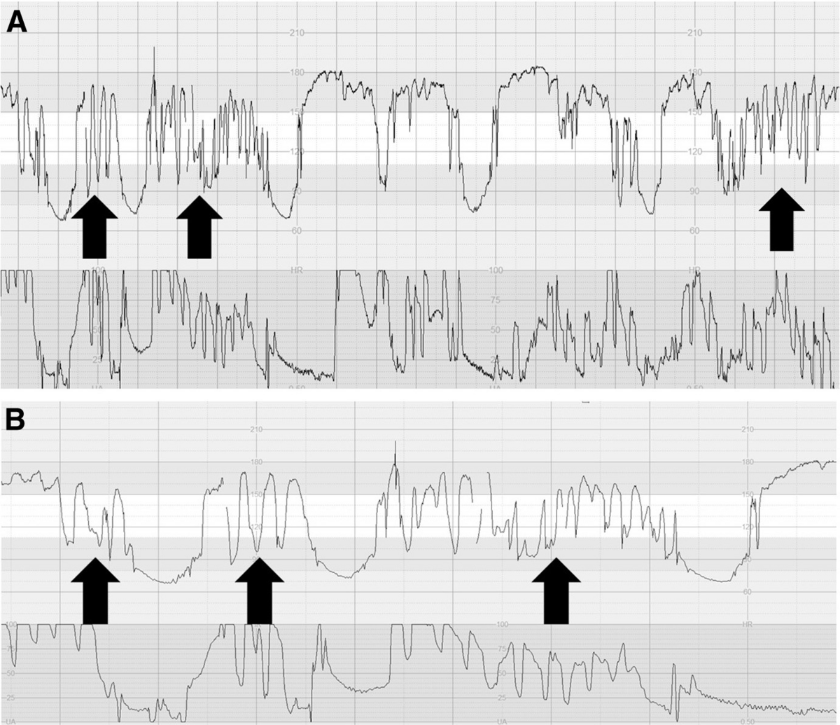 Significance: a ZigZag pattern lasting > 2 min is associated with an ~11-fold increase in neonatal-unit admission, and > 1 min with a ~2-fold increase in neonatal acidosis. When it accompanies an increased baseline FHR without repetitive decelerations, it should raise the suspicion of fetal neuroinflammation (chorioamnionitis). A ZigZag pattern persisting > 1 min requires immediate action to improve fetal oxygenation (reduce/stop oxytocin, consider tocolysis; stop active pushing if seen with a subacute hypoxic pattern). 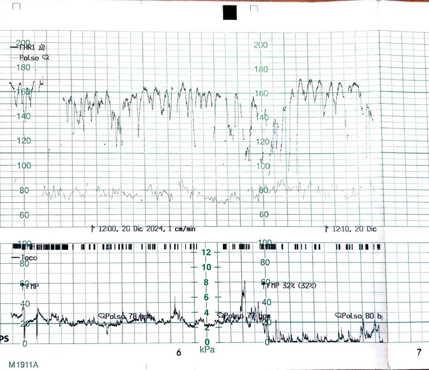 [1] [2] [3]
|
| Saltatory |
Saltatory pattern (FIGO "increased variability").
A uniform increase of the baseline bandwidth exceeding 25 bpm that lasts for more than 30 minutes. 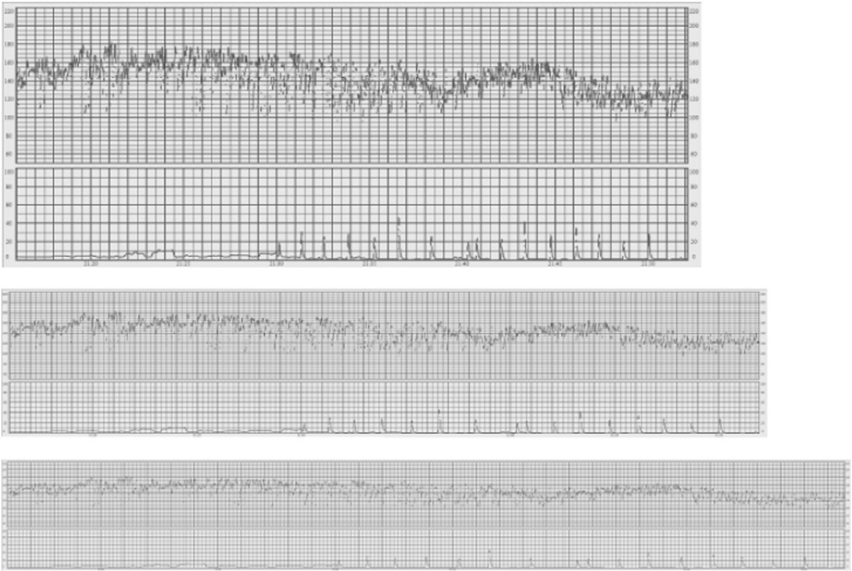 A true saltatory pattern (> 25 bpm for > 30 min) is rare (< 5%) during labour: ongoing uterine contractions intermittently interrupt fetal oxygenation, so a uniform increase of the bandwidth usually cannot be sustained for 30 min. When present, it is mostly seen antenatally, reflecting recovery from an acute and profound (non-fatal) hypoxic-ischaemic insult, and is attributed to autonomic CNS instability. During labour, an erratic (non-uniform) fluctuation of > 25 bpm should instead be classified as a Zigzag pattern. [1] [2] [3]
|
| Sinusoidal pattern |
Sinusoidal pattern — a regular, smooth, sine-wave-like oscillation of the baseline FHR, amplitude about 5–15 bpm (a broad, lower-amplitude definition down to ~2–5 bpm may be used when the clinical context fits), frequency 2–5 cycles per minute (classically 3–5), lasting at least 10 minutes, with absent baseline variability and no accelerations.
Pathophysiology: classically severe fetal anaemia (anti-D alloimmunisation, feto-maternal haemorrhage, twin-to-twin transfusion, ruptured vasa praevia); also described in acute fetal hypoxia, infection, cardiac malformation, hydrocephalus and gastroschisis. This annotation currently groups both sinusoidal sub-types — they are not separated for the challenge, for lack of data (sub-annotation remains possible when the information is available, cf. issue #11): Typical (smooth, rounded, symmetrical sine wave; Graça type I): seen in fetal anaemia, may occur antenatally. 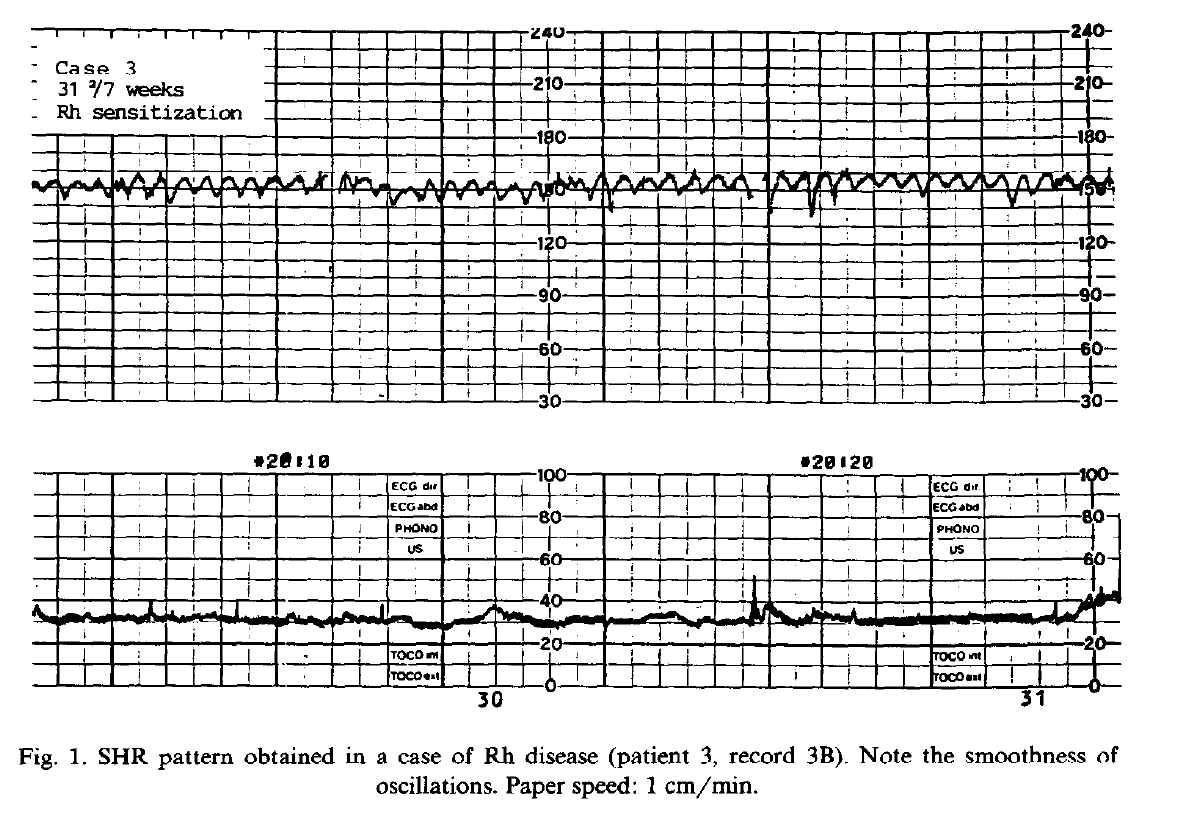 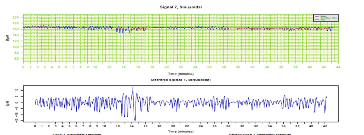 Atypical — jagged / saw-tooth, the “Poole Shark Teeth” pattern (Graça type II): secondary to sudden feto-maternal haemorrhage, almost always intrapartum; usually mandates immediate delivery. 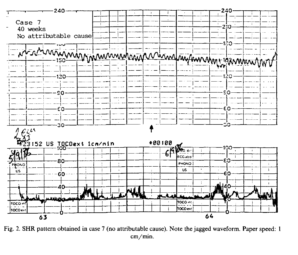 Distinguish from the pseudo-sinusoidal pattern (jagged, short-lived < 30 min, normal patterns before/after, benign clinical context) and from the saltatory pattern. [1] [2] [3] [4]
|
| Not analysed/Unsure |
Not analysed / Unsure (variability) — a baseline segment whose variability has not been annotated or cannot be classified with confidence. Excluded from the variability ground truth.
|
| General |
A window of interest is a window that the AI can see to analyse the signal. It should include all possible useful periods to properly analyse what is annotated.
Default: 10 min after/before the last/first annotation. |
| Force in window of interest |
Force in window of interest — mark a segment so it is forced into the window of interest: it is kept as part of the data made available to the artificial-intelligence model even if not annotated.
|
| Not in WOI |
Not in window of interest — a portion of the recording that lies outside the window of interest and will not be used for AI training / evaluation.
|
| Clear |
Clear — an eraser: applied over a period it removes any existing annotation there, leaving the trace blank / unannotated (it does not itself create a labelled period).
|
| Window of interest |
Window of interest (WOI) — the portion of the recording that is used for AI training / evaluation. By default it is computed automatically around the annotated events (with a few minutes of margin); it can be overridden with Force in WOI.
|